RapiClear
顯微樣品透明液
RapiClear是一種水溶性透明試劑, 用來將生物組織迅速且輕易的透明化. 它可以加強已標定螢光染劑
樣本的視覺化深度達 um 甚至到 mm 等級。
RapidClear已被廣泛運用於動植物、昆蟲等細胞型態觀察, 同時用於膠原蛋白、纖維素等生物物質支架
的描繪, RapiClear的應用促使生物樣本細部3D影像之建構成為可能 !
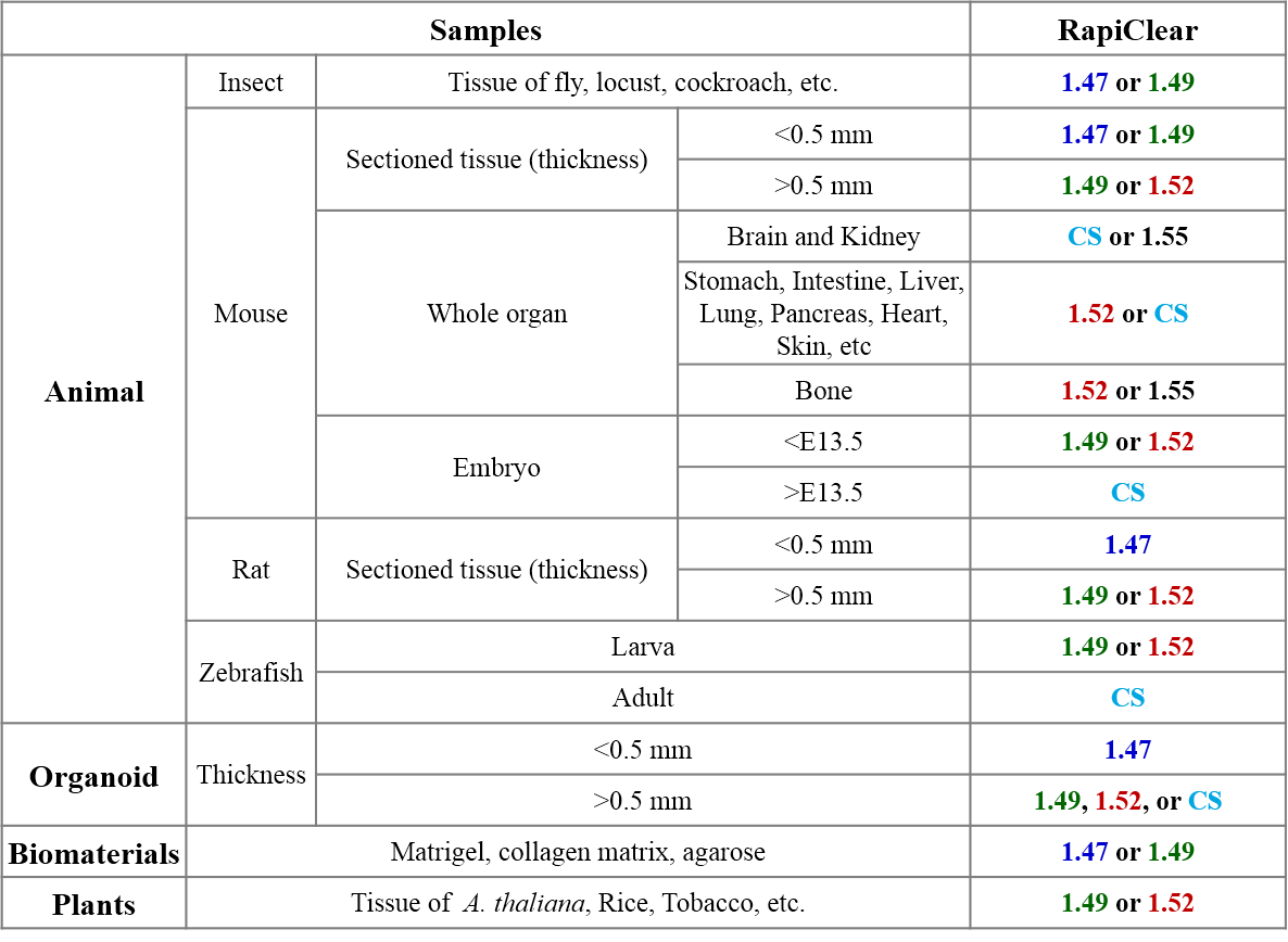
RapiClear 選擇參考表 (for different samples/tissue)
Multiscale 3D Image
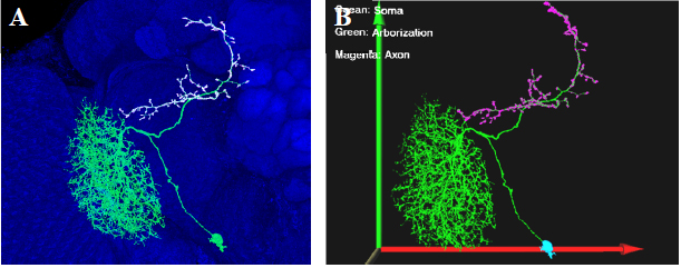
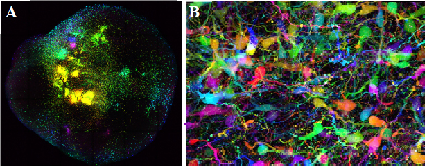
圖一 Fly brain in RC147 A. Expression pattern of a Drosophila optical lobe projection neuron. Neuron arborization is labeled by mCD8::GFP (green), pre-synaptic terminals are labeled by anti-synaptotagmin::HA Ab (white), and brain structure are counterstained by anti-DLG immunostaining (blue). B. 3D visualization of the optical lobe projection neuron showed in figure A .
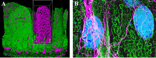
圖二 Mouse intestine and pancreas in RC152 A. Mouse intestine. Blood vessels in magenta (lectine) and nuclei in green (Sytox). B. Mouse Islets. Blood vessels in green (lectine), islets in blue (insulin) and nerves in magenta (TUJ1).
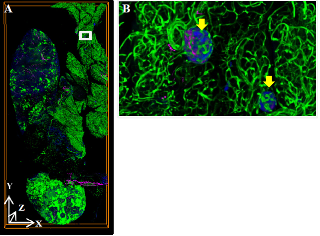
圖三 Mouse pancreas in RC152 A. Map derived from 55 tile-scanned images. Magenta: neuronal staining. Blue: insulin staining. Green: vascular staining. X-axis, 6mm; Y-axis, 13mm; Z-axis, 0.5mm. B. Magnified view of islets in pic. A (box) Yellow arrows indicate islets.
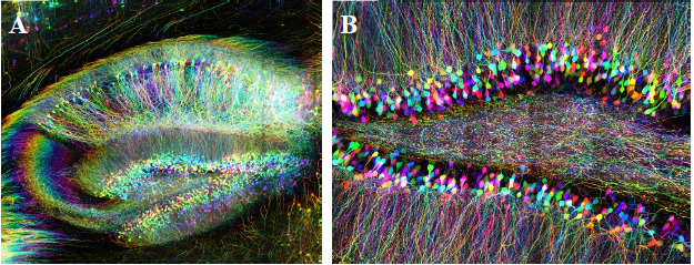
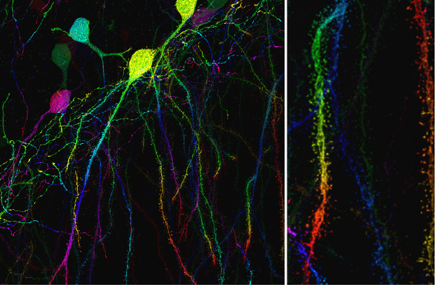
圖四 Mouse brain in RC149 and RC152 Depth coded projection images show YFP-expressing hippocampal neurons in 550um mouse brain slices. A. Sample cleared with RapiClear 1.49 and imaged with 25x lens. B. Sample cleared with RapiClear 1.52 and imaged with 63x oil lens.
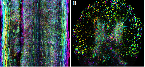
圖五 Mouse spinal cord in RC149 Depth coded projection images show YFP-expressing neurons in RapiClear 1.49 cleared 550um mouse spinal cord slices. Imaged with 25x lens. A. Sagittal section. B. Coronal section.
圖六 Mouse muscle in RC149 and RC152 Depth coded projection images show YFP-expressing neurons in mouse gastrocnemius muscle slices. A. 1000 um slice cleared with RapiClear 1.49 and imaged with 10x lens. B. 550 um slice cleared with RapiClear 1.52 and imaged with 63x oil lens.
圖七 Organoid in RC149 GFP-expressing neurons from a 1.2mm cerebral organoid cleared with RapiClear 1.49 are shown in depth coded projection images. A. imaged with 10x water lens. B. Magnified view in pic. A under the 63x oil lens.
圖八 Mouse brain in RC152 Depth coded projection images show YFP-expressing hippocampal neurons in mouse brain slices imaged with super-resolution microscopy.
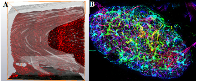
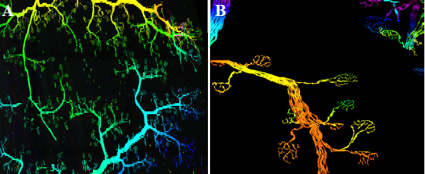
圖九 Mouse long bone and lymph node in RC152 A. Mouse long bone. Fibrillar collagen in gray (SHG) and bone marrow in red (DsRed). B. Depth coded projection image shows the blood vessel circuits in the whole lymph node.
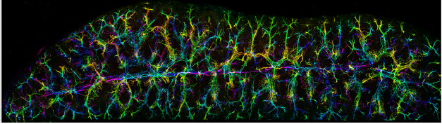
圖十 Mouse spleen in RC152 Depth coded projection image shows the nervous system in 550um mouse spleen slice.
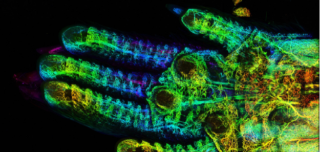
圖十一 Whole mouse paw in RC152 Depth coded projection image shows the blood vessel circuits in the mouse paw.
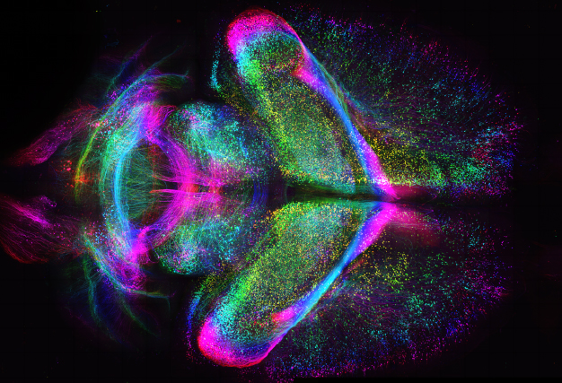
圖十二 Whole mouse brain Depth coded projection image shows the Thy1-GFP mouse brain neuronal circuitry cleared with CLARITY-RapiClear 1.47.
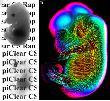
圖十三 Whole mouse embryo A. The transmitted white light images show before and after CLARITY-RapiClear optical clearing. B. Depth coded projection image shows the E13.5 mouse embryo neuronal circuitry labeled with anti-TUJ1 antibody and cleared with RapiClear 1.47 reagent.
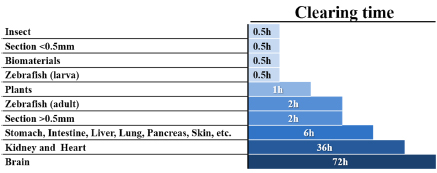
RapiClear對不同樣品的光學透明化速度: